Postingan

Menampilkan postingan dengan label Pleural Effusion

7 Examination of Pleural Effusion
Gambar
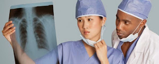
1. Chest X-rays Chest X-rays are usually the first step for diagnosing pleural effusion, the results of which indicate the presence of fluid. Surface of the liquid contained in the pleural cavity will form a shadow-like curves, the lateral surface area is higher than the medial surface. When the horizontal surface of the lateral to medial sure the air contained in the cavity that can come from outside or inside the lung itself. Another thing that can be seen in the photograph chest, mediastinal pleural effusion is classified on the opposite side of the liquid. However, if there is atelectasis on the same side with the fluid, mediastinal will remain in place. 2. CT scan of the chest CT scan clearly depicts the lungs and fluid and can indicate the presence of pneumonia, lung abscess or tumor. 3. Ultrasound chest Ultrasound can help determine the location of the collection of small amounts of fluid, so that the discharge can be done. 4. Thoracocentesis Aspiration of pleural fluid is usefu...

Nursing Care Plan for Pleural Effusion

Gambar
Pleural effusion Pleural effusion is excess fluid that accumulates in the pleura, the fluid-filled space that surrounds the lungs. Excessive amounts of such fluid can impair breathing by limiting the expansion of the lungs during respiration. Etiology of Pleural Effusion Various causes of pleural effusion are: Neoplasms, such as bronchogenic and metastatic neoplasms. Cardiovascular, such as congestive heart failure, pulmonary embolus and pericarditis. Diseases of the abdomen, such as pancreatitis, ascites, abscess and Meigs syndrome. Infections caused by bacteria, viruses, fungi, and parasites microbacterial. Trauma Other causes such as systemic lupus erythematosus, rheumatoid arthritis, nephrotic sindroms and uremia. Signs and Symptoms of Pleural Effusion Cough Dyspnea varies Complaints of chest pain (pleuritic pain) In severe effusions occur protrusion intercostal space. Chest movement was reduced and delayed on the part of the experience effusion. Percussion dims above pleural eff...